Magnetic nanoparticles can be used for medical imaging, to isolate disease markers, to transport drugs through the body and even to burn away cancer cells. Randy De Palma and Gustaaf Borghs of IMEC report on a new procedure to efficiently coat magnetic nanoparticles, tuning their properties to the doctor's needs
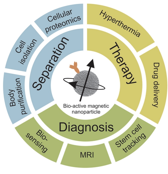
The ability to make magnetic particles the size of biological functional units, such as proteins, enzymes or DNA, has opened up new ways to interact with the biological world. By making these magnetic nanoparticles water-soluble and coating them with suitable biological receptors, their unique nanoscale magnetic properties can be further exploited to allow for highly effective biomedical separation, diagnosis and therapy (Figure 1). When functionalised with the proper receptors, magnetic nanoparticles can specifically interact with biological targets, such as cells (e.g. tumour cells), organelles, viruses, biomolecules, etc.
A well-known application in the field of diagnosis is the use of magnetic nanoparticles as next-generation imaging probes for observing biological events via magnetic resonance imaging (MRI). Although MRI is one of the most powerful medical diagnostics tools due to its non-invasive nature, high spatial resolution and multidimensional tomographic capabilities, it lacks in sensitivity.
This weakness can be overcome by using magnetic nanoparticles as contrast agents. MRI is for example used to better differentiate healthy and pathological tissues. Due to their low toxicity, iron oxide magnetic nanoparticles have received a Food and Drug Administration (FDA) approval to be used as MRI signal enhancers. Another diagnostic application is the magnetic labeling of stem cells to monitor the fate and distribution of transplanted stem cells non-invasively in the human body.
Furthermore, magnetic nanoparticles show great promise as magnetic labels in biosensing with many advantages compared with the conventional labels such as enzymes, fluorescent dyes, chemiluminescent molecules or radioisotopes.
Next to their small size and low toxicity to humans, magnetic nanoparticles have the advantage that they are subjective to an external magnetic field gradient, which moreover can penetrate deep into the human tissue. This property has inspired the medical world to various therapeutical applications. A first example of nanoparticle-based therapy is the controlled transport of drugs to target sites.
The latter is realised by attaching a drug to a biocompatible magnetic nanoparticle carrier, injecting the ferrofluid into the bloodstream and applying an external magnetic field to concentrate the drug/carrier complexes at the target site. This principle is for example used with cytotoxic drugs in cancer treatment. Another interesting therapy is based on the magnetic nanoparticles ability to be heated when a time-varying magnetic field is applied. This characteristic is used to burn away cancer cells (hyperthermia), often in combination with chemotherapy. It is in fact known that cancer cells are more sensitive to temperatures in excess of 41°C than their normal counterparts. Both applications present a bright future for targeted therapy, which allows specifically destroying a desired target without deteriorating healthy surrounding tissue.
Finally, the attraction between an external magnet and the magnetic nanoparticles enables separation of a wide variety of biological moieties. Examples are the isolation of cancer cells in blood samples or stem cells in bone marrow to allow for improved diagnosis and the removal of toxins from the human body. Furthermore, magnetic nanoparticles can be biologically activated to allow the uptake of cells via endocytotic pathways. By this means, certain cellular compartments can be specifically addressed. Once taken up, the desired cellular compartments can be magnetically isolated and accurately studied using proteomic analysis.
There exist many synthesis protocols for magnetic nanoparticles, based on micro-emulsion, coprecipitation and other water-based methods. The disadvantage of these methods is that the size uniformity and crystallinity of the magnetic nanoparticles is rather poor. Recently, Sun et al. developed a new and simple synthetic procedure that enables high-quality ferrite magnetic nanoparticles with sizes between 3 and 20 nm. A typical process involves the high-temperature decomposition (> 220°C) of an organic iron precursor in the presence of hydrophobic ligands such as oleic acid. This method has been further adapted by other researchers for the synthesis of all types of magnetic nanoparticles containing different materials such as Cobalt, Manganese, Nickel, Platinum, etc (Figure 2).
Although this thermal-decomposition method has the advantage of producing very monodisperse particles, it has the big disadvantage that the resulting nanoparticles are only soluble in non-polar solvents due to their coating with hydrophobic ligands. Hence, to make magnetic nanoparticles suitable for biological applications, the hydrophobic ligand coating needs to be replaced by a hydrophilic, biocompatible and functional coating that allows controlled interaction with biological species.
Ligand exchange can be used to replace the hydrophobic coating of the abovementioned magnetic nanoparticles with hydrophilic molecules (Figure 3). This method involves adding an excess of ligand to the nanoparticle solution, which results in the displacement of the original ligand on the nanoparticles' surface. In this way, research groups have realised ferrite magnetic nanoparticles covered with hydrophilic ligands containing carboxylate, phosphate and alcohol end groups, making the particles water-soluble. However, the long-term stability of these water-soluble nanoparticles has not been unambiguously proven due to the weak (non-covalent) binding of the ligands to the magnetic nanoparticles.
Based on its expertise in silane self-assembled monolayers for biosensor applications, IMEC recently devised a procedure to coat magnetic nanoparticles (realised via the thermal-decomposition method) with silane monolayers. The advantage of this approach is that it enables stable, water-soluble monodisperse magnetic nanoparticles bearing a huge variety of functional end groups. The latter makes it possible to tune their surface functionality, optimising it for every specific application.
IMEC performed a systematic study of silane ligand exchange, screening nine commercially-available silane monolayers, i.e. amino-silane, short aldehyde-silane, isocyanate-silane, thiol-silane, cyano-silane, acrylate-silane, long aldehyde-silane, carboxylic acid-silane and poly(ethylene glycol)-silane (Figure 3). A first observation - made for all silanes - was that the original hydrophobic ligand was fully replaced by the silane self-assembled monolayer. The successful silane ligand exchange was confirmed using X-ray photoelectron spectroscopy and Fourier transform infrared analysis.
The silanes formed a dense organic layer with the functional end groups presented to the surrounding liquid. Furthermore, it was shown that the presence of these endgroups strongly determines the water-dispersibility of the nanoparticles. Amino, carboxylic acid and poly(ethylene glycol) were found to render these nanoparticles soluble in aqueous solutions over a wide pH range (Figure 4).
In general, these coatings can be described as being respectively positively charged, negatively charged and neutral (Figure 5). It was recognised that surface hydrophilicity, in combination with electrostatic and steric repulsions, play an important role in the stabilisation of MNPs.
After silane ligand exchange the hydrodynamic diameter increased from ~9 nm up to ~13 nm and the magnetic character per nanoparticle was not affected. Compared with other ligands commonly used to stabilise ferrite MNPs, an enhanced long-term stability was observed and an increased resistance against mild acid and alkaline environments. This was dedicated to the covalent linkage of the silane layers onto the nanoparticles' surface.
The combination of Sun's thermal-decomposition method to manufacture high-quality magnetic nanoparticles and IMEC's procedure to coat the nanoparticles via silane ligand exchange is a true success formula. It represents a generic and versatile method to synthesize highly stable, water-soluble ferrite MNPs with a variable ligand periphery and tunable surface properties.
Future work will among others focus on the use of magnetic nanoparticles for endosomal proteomics that can for example contribute to a more fundamental understanding of the biochemistry behind Alzheimer disease. The endocytotic uptake of magnetic nanoparticles will be studied as a function of the nanoparticles surface functionality, which can be easily tuned using the above-mentioned silane chemistry. Following uptake by the cells, the cellular endosomal compartments are magnetically labeled and can be effectively isolated and studied using proteomic analysis.
These endosomal compartments contain crucial information to understand the disease pathology. Since a wide range of silane molecules is commercially available, one can easily screen the effectiveness of the available end groups for a specific application, ranging from cell isolation, over medical imaging, to hyperthermia for cancer treatment.


