More weapons are needed to stop counterfeit tablets entering the market. Dr Detlef Beckers, at PANalytical BV, describes how X-ray powder diffraction can detect counterfeit tablets in blister packs without even opening them
X-ray powder diffraction (XRPD) is well established as a key analytical tool in the pharmaceutical industry. Now, new developments, such as ultra-fast X-ray detectors, make the technique a powerful tool for quality control. Applications include monitoring for structural changes that can occur during production, packaging or storage, and for policing and identification of counterfeit drugs.
Counterfeiting is a growing concern: Annual earnings from counterfeit drugs are estimated to be between US$20 bn and $50 bn (Euro 12.6-31.5bn). The US Center for Medicines in the Public Interest forecasts that sales could reach $75 bn ( €47.3bn) in 2010. Such high financial rewards inevitably attract organised criminal groups that are willing to invest in increasingly sophisticated technologies to avoid detection and protect profits.
The internet serves the counterfeit trade as a valuable distribution channel and communication network at a global scale although the greatest financial returns can be achieved by penetrating pharmaceutical supply chains at the national level. Today, counterfeit drugs are frequently being detected even in countries with highly regulated pharmaceutical supply chains. For example, in the UK the Medicines and Healthcare products Regulatory Agency states that counterfeit medicines have entered the regulated supply chain and reached patient level on nine occasions over the past three years.
In March 2008, the European Commission launched a public consultation in preparation for a legal proposal to combat counterfeit medicines. Welcomed by the European Pharmaceutical Industry Association, the move should result in regulatory reform.
While the source of the counterfeiting problem must be addressed with economic and regulatory measures, it is also important to look at methods that aid discrimination of fake drugs from real ones. In many cases the cardboard cartons, blister pack and foil backing, holograms and logos, as well as the appearance of counterfeit drugs, are so well copied that there is no visual difference between fake and authentic products.
Similarly, shelf-life studies of drug substances in commercial dosage forms are an essential element of the QC process for manufactured pharmaceuticals. Good Manufacturing Practice (GMP) requires the product to have a proven shelf-life with minimal levels of decomposition of the active ingredient that would reduce the potency or cause the formation of impurities that might be harmful to the patient.
To date, reliable differentiation has required the application of analytical techniques following removal of the tablet from the blister. However, once taken from its original packaging the drug cannot be sold.
Now, a testing method using the unique "fingerprinting" mechanism offered by XRPD provides the opportunity to analyse tablets without requiring removal from their blister packaging.
X-ray powder diffraction (XRPD) is a rapid, versatile technique that reveals detailed information about the chemical composition and crystallographic structure of natural and manufactured materials. XRPD allows, for example, qualitative and quantitative phase analysis of the various constituents of the complete solid drugs, including the active pharmaceutical ingredient (API) and the fillers, with very low detection limits and control of the final dosage form. Importantly, the technique is non-destructive; samples are left intact and can be re-analysed or used in other processes. Applications include a variety of research and industrial uses.
A crystal lattice is arranged to form a series of parallel planes separated from one another by a distance (d), which varies according to the nature of the material. For any crystal, planes exist in a number of different orientations each with its own specific d-spacing.
The phenomenon of X-ray diffraction by crystals results from a process, in which X-rays are scattered by the electrons of the atoms in the material. When a monochromatic X-ray beam with wavelength lambda (l) is projected onto a crystalline material, diffraction occurs only for those angles theta (q) where the conditions of the so called Braggs law [nl =2dsinq] are fulfilled. The intensities of the diffraction maxima are related to the strength of those diffractions in the specimen; they depend on the nature and quantity of the material.
Plotting the angular positions and intensities of the resultant diffracted peaks of radiation produces a pattern, which is characteristic of the sample - like a fingerprint. Where a mixture of different phases is present, the resultant diffractogram is formed by addition of the individual patterns. Therefore phase identification can be accomplished by comparing the data (peaks and relative intensities) from the specimen with peaks and relative intensities from databases.
In transmission X-ray diffraction experiments, the incident X-ray beam is not reflected by the sample, but instead travels through the sample, where the diffraction process takes place.
Materials consisting of light atoms, such as organic compounds in pharmaceuticals, are relatively "transparent" for X-rays. In the transmission geometry the X-ray beam, coming off the focus of the X-ray tube, is typically focused on the detector by an incident beam conditioner - an X-ray mirror, for instance.
In contrast to a crystalline pattern consisting of a series of sharp peaks, amorphous materials (liquids, glasses and most polymers used in pharmaceutical packaging) produce a broad background signal although some polymers do show semi-crystalline behaviour.
A study was designed to demonstrate the power and flexibility of XRPD analysis, with the ability to distinguish polymer and metallic packaging materials from the pharmaceutical compounds held safely within it.
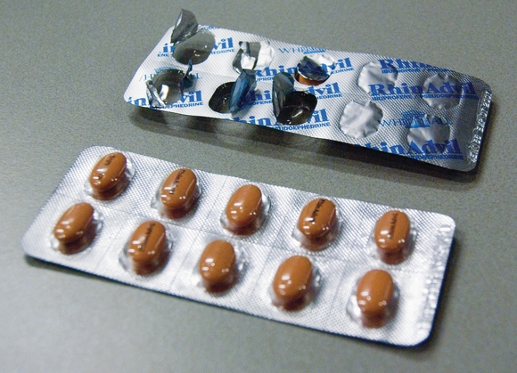
The tablets studied are commercially available from European pharmacies or specifically produced tablets for testing purpose. All were contained within blister packs with a polymer cavity and aluminium lidding.
XRPD measurements were performed with a PANalytical X'Pert PRO MPD diffractometer, equipped with a focusing mirror and motorised high-throughput stage. The blister cards were mounted on a metal frame and the diffraction signals were measured in transmission mode using an X'Celerator detector.1
XRD measurements on emptied blisters: XRD measurements were made on empty blister packs to determine any packaging effects. Results showed that the amorphous polymer cavity caused an increased, modulated background, while the aluminum lidding produced some intense, easily identifiable Bragg peaks at high angles, confirming that the packaging material should not interfere significantly with the unique fingerprint of tablets.
To determine the intensity attenuation effect of blister packaging, 10 different commercial blister cards were investigated, including transparent and opaque types, with aluminium or aluminium/paper laminate. All packaging types showed only a moderate attenuation effect (absorption factors mostly in the range of 2.5 to 3.5), confirming the ability of X-rays to "see through" the packaging.
XRPD measurements of tablets through the blister: Figure 1 shows XRPD data from a single commercial tablet in an opaque blister and the signal from the emptied blister. The presence of the packaging material is evidenced mainly by an increased background and by the two Bragg peaks at high angles. This shows that high quality diffraction data can be obtained directly from the intact, blister-packed tablets, even with opaque blister materials.
Automated blister card scanning: To demonstrate high-throughput scanning, a 3 by 6 blister card was analysed. Measurement time for each tablet was 10 minutes (three hours for the whole card). Results showed that a few tablets differ in some details from the others (Figure 2). This could be due to differences in preferred orientation, particle statistics or composition. It was not the intention of this study to determine the reason for these discrepancies, however the data serve to illustrate that the method is fully capable of identifying tablets that differ from the norm.
Detection of fake drugs or polymorphic changes of tablets in a blister pack: Tablets with different compositions were prepared in blister packaging to simulate drugs containing different or no API; or tablets where polymorphic changes have occurred (Table 1). Tablets were placed in an empty commercial blister card and carefully resealed.
Samples were measured in automated mode. Figure 3 shows that differences in crystal form and/or API amount are clearly observed, mainly through the absence or presence of diffraction peaks at angles below 25Ëš.
Identification of counterfeit drugs: Figure 4 shows the diffraction patterns of an original tablet and a counterfeit copy product. Both tablets contain the same amount of API, but the diffraction patterns are clearly different showing a difference in crystallinity of the API (causing an expected difference in the bioavailability) as well as differences in the composition of excipients in the tablets.
in Summary, XRPD analysis of tablets in intact packaging is a non-invasive technique that provides high quality information on the structure and composition of materials. Analysis can now be conducted, through even fully opaque blister packs. This allows effective, quick and reliable screening. High throughput is achievable with latest generation systems. This innovative method for monitoring drug quality can be used to differentiate between authentic drugs and counterfeit products.


