In recent years, second-harmonic generation (SHG) microscopy has established itself as the technique of choice for the study of crystallised biomolecules such as starch, collagen and myosin.
Unlike fluorescence-microscopy, SHG microscopy does not involve the creation of excited electronic states, so cell viability issues associated with heating and photobleaching are reduced.
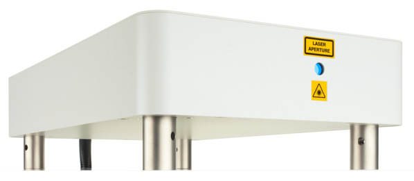
The report describes typical results from collagen in liver tissue samples generated by SHG microscopy using a Chromacity 1040 laser as the excitation source.
In these experiments, this laser is shown to be an ideal source for an SHG microscopy system as it allows researchers to generate exceptionally clear, high-resolution images because of its excellent beam quality, ultra-fast pulses, and high average power levels.
Further by operating at near infrared wavelengths, the Chromacity 1040 enables the construction of 3D images of specimens by imaging deeper into thick tissues.
Design optimised to operate at 1040 nm, SHG microscopy using a Chromacity 1040 laser enables the direct visualisation of tissue structure (in situ) as it relies only on species present in the sample to provide a contrast.


